


Project DIG was a partnership between Queensland Museum, BHP and BMA to transform how we store, explore and share our collections and research with communities worldwide.
From dig to digital - Bringing specimens to life in 3D
About
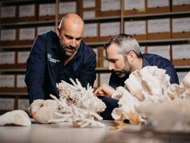
Museums worldwide have a big problem that needs a big solution. Museums hold the world’s most precious treasures, from celestial meteorites, to tiny fossils smaller than a grain of sand. Millions of specimens in these collections tell the grand story of our Earth, its past, its present and each specimen holds the clues to a future Earth.
Museum scientists alone cannot study every specimen in the collection, nor can museums put every specimen on display. With lifetimes of research needed, across countless experts much of a museum collection remains under-studied. Much of the collection is stored away from public view simply because these specimens are physically too hard to display. Some are so large, they represent entire sites, or so small the human eye cannot see them or simply too fragile that even exposing them to the air or light can be their destruction. Museums worldwide are working to solve this problem through cutting-edge digital technologies.
Project DIG is working to solve this big problem for Queensland Museum by developing some big solutions, to take the museum’s collections from behind closed doors and share this with the scientific community and the public.
Our researchers are combining new scanning technologies, such as photogrammetry, structured-light scanning, and X-ray computed tomography (CT), along with 3D modelling and LiDAR to capture museum specimens (inside and out), at every point of their journey from their discovery in the field to scientific study and display. Each digital model, called a cybertype, represents a unique digital insight into the specimen and provides opportunities for study globally and through ways, the physical world cannot provide.
Constructing a complete picture of life in ancient Queensland
Digital Replicas: capturing 3D reality through photographs, light and lasers
Museums have produced replicas of the real world for hundreds of years. Museums used to (and still do) cast dinosaur bones, or even an entire archaeological sites, using silicon rubber moulds filled with plaster and then painted. Unfortunately, this analogue process does not work for huge sites or intricate and fragile specimens. The traditional methods also introduce foreign materials and chemicals like silicon rubber, plasticine, latex and plaster into the specimens that are being replicated. However, digital 3D gets around all of this by using hands-off techniques to produce a digital replica. Museums are no longer bound by the realities of the physical world.
Our researchers use pixels, light, and lasers to capture the surface of sites, specimens and objects in a range of contexts, without needing to use any replication materials. These techniques include drone photogrammetry and LiDAR in the field through to micro-photogrammetry and light surface scanning in the lab. Each technique provides the museum with different abilities to capture the real world in virtual in detail never before thought possible.
Photogrammetry
Photogrammetry is not a new technology, but digital photogrammetry is and has revolutionised the 3D capture of the real world. Using hundreds to thousands of individual overlapping 2D digital photographs, captured from around a site, object or specimen, software can turn millions of two-dimensional pixels into three-dimensional points in space (a photogram model). This software process extracts the 3D pixels and produces a 3D ‘point cloud’. The cloud of points reflects the ghostly shape of the real subject captured in the photos. The more points you have the more detailed your cloud becomes, and the better your final 3D model. The software then creates a surface between these 3D points, turning what looked like a dust cloud into a solid object! These models can be used for all sorts of research, animation, display and even 3D printing.
Drone (UAV) photogrammetry
Fossil sites in Queensland are often very remote and can be difficult to access. Or sometimes, these sites might be under threat loss. Each time you excavate a fossil site you change it forever. So our scientists capture these sites before they dig, “digitise before you dig!”. Drones, or unmanned aerial vehicles, fly around very large sites, from 10s of meters to 100s of meters in size, photographing the environment as it goes. These photos are stitched together to create a 3D model of the site before and after it has been excavated, producing a 3D model of the fossil site through time (4D). With all of this 3D data, anyone in the future can re-visit a site and see what it looked like before it was changed. In the future, 4D scans will be how we ‘time-travel’ back to places and things in the past to see what they looked like!
Micro-photogrammetry
Back at the lab, our palaeontologists use super high definition macro-lens cameras to capture our smallest specimens, only millimetres in size. As the specimens rotate on a turntable, they are captured in 3D, and using the same process of photogrammetry, models of each specimen are produced in micron accuracy. Tiny fossils need to be looked at under a microscope but they are exceptionally fragile and even the slightest bump could break them. Each time a scientist moves the specimen to study them, they risk damage to an irreplaceable specimen. High resolution 3D models of these tiniest specimens reduces the need to handle our precious specimens, assisting with their long term preservation.
Structured Light surface scanning
Much of a museum’s collection is dedicated to ‘Type Specimens’. Type Specimens are the most important representatives of any extinct or living species in existence and need to be preserved and conserved in perpetuity (over hundreds of years). However, scientists need to be able to see these specimens for their research. In the past, specimens may have risked being lost through postage or risked damage by trying to replicate them. Instead, high resolution surface scans can provide these researchers with enough detail to allow them to compare their specimens against others around the globe. We use micron-resolution structured-light surface scanning to help produce these models in an automated way. Specimens are placed on a turntable that automatically moves and predicts which directions the specimen needs to be positioned to scan. Structured-light scanning works in tandem with colour photogrammetry to produce the best resolution for a researcher to study.
Scanning technology in palaeontology
X-ray visions reveal new worlds in old bones
Photogrammetry, structure-light and laser scanning all are used to produce excellent 3D models of the surface of a site, object or specimen. But what if you want to look inside it? Many fossils are trapped within rock that obscures it, or there are features within the body of an animal that researchers would like to examine such as bones, organs or even mummies. X-rays allow scientists to see what is inside something without having to cut it open and potentially destroy it.
Medical imaging technology has advanced to help people (and pets) to see if there's something internally that might be making them ill. Computed Tomography (CT), or CAT scanning, is a technology that pieces together thousands of individual X-ray pictures into a three dimensional image. Museum scientists discovered that using the same X-ray technology they can look through a variety of specimens and objects, from dinosaur bones and eggs, to animals and plants, Egyptian mummies or even look inside at the piece of a mobile phone.
In palaeontology we use X-rays to see if we can look for fossils before treating them with chemicals or using tools to chip away at rock. There might be features we cannot see with the naked eye, like a hatchling dinosaur, or footprints buried inside a rock, or minerals growing within a meteorite. Without X-rays, the only other way to achieve this would be to slice a specimen into tiny slivers, looking at each one piece by piece, destroying the fossil in the process.
Revealing hidden bones using scanning technology
CT scanning dinosaur trackways
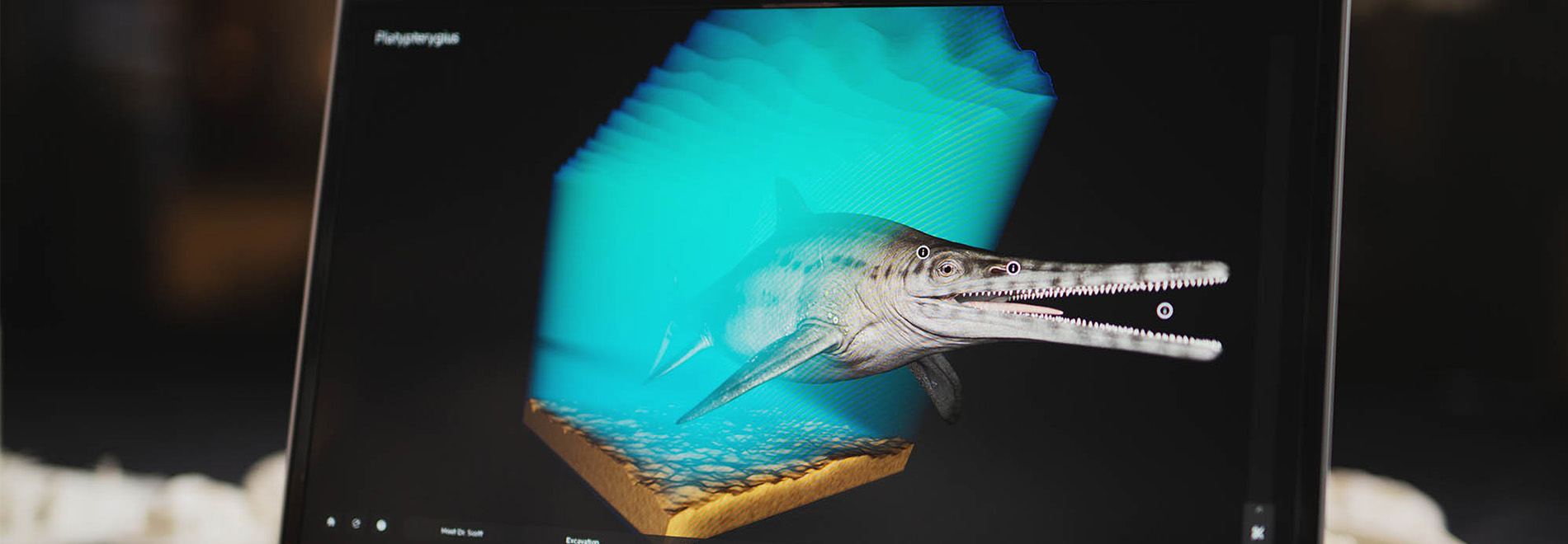
Exploring real scientific data
Many 3D digital experiences available to the public are fiction. Whether this is an online game or an animation created by artists with only concepts to use. Authentic museum experiences need to use the original 3D model created directly from its collection items and sites.
Project DIG has partnered with Swedish based 3D-visualisation software company Interspectral to transform our collections and research into amazing digital exhibitions. Inside Explorer software takes real scientific datasets and composites them into a story about the specimen and the environment around it. Visitors can interact with our collections and research through touch screens and touch tables providing an engaging learning experience for people of all ages.
Digital exhibitions provide greater public accessibility to our collections, allowing our visitors to engage and interact with our rare, fragile and valuable specimens.
Creating digital exhibitions using real scientific data
Images
1 of 5
You might be interested in
Projects
Find out more about some of our long-term Collections & Research projects.

Project DIG
Discover how Project DIG unlocked knowledge held in our State Collection for visitors and researchers worldwide.

Research projects
Project DIG delivers innovative research projects that allow digital exploration, revealing new and exciting information for scientists, experts and leaders worldwide.